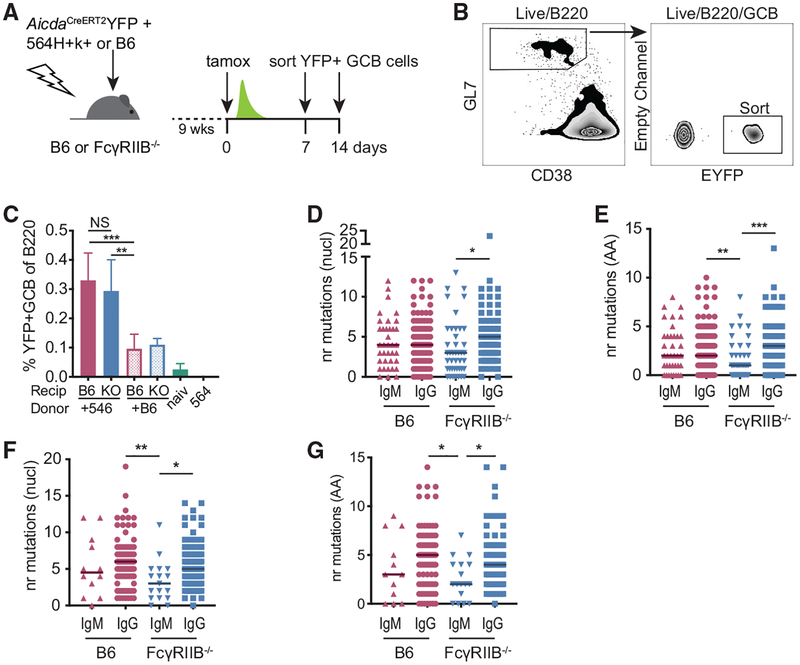
Figure 6. Low SHM IgM+ GC B Cell Clones Persist in the Absence of FcγRIIB on FDCs.
(A) Experimental setup to label and follow 564Igi-induced, autoreactive GC B cells. B6 or FcγRIIB−/− mice were lethally irradiated and subsequently received a 1:2 mixture of 564Igi and AicdaCreERT2EYFP BM or a 1:2 mixture of B6 and AicdaCreERT2EYFP BM. After 9 weeks, mice were tamoxifen induced (tamox) and splenic EYFP+ GC B cells were sorted at days 7 or 14 post-tamoxifen.
(B) Gating strategy used to sort EYFP+ GC B cells. Cells were gated on forward versus side scatter (FSC/SSC) for lymphocytes, singlets, and live-dead stain negative cells before the indicated gates.
(C) Frequency of EYFP+ GC B cells relative to measured B220+ B cell population in indicated chimeras and naive AicdaCreERT2-EYFP mice (naiv) and 564Igi mice at the 1 week post-tamoxifen time point. Recip, recipient; KO, FcγRIIB−/−. All of the chimeras received 2 parts AicdaCreERT2-EYFP (not shown) and 1 part 564Igi (+564) or 1 part B6 (+B6). Pooled data from 3 independent experiments. n = 6 mice for 564Igi+AicdaCreERT2 EYFP chimeras, 4 mice for B6+AicdaCreERT2EYFP recipients, naive AicdaCreERT2EYFP, and 564Igi mice. Error bars represent SDs.
(D and E) The number of nucleotide mutations (D) or amino acid mutations (E) in the VDJ region from EYFP+ GC B cell heavy chains obtained from 564Igi+AicdaCreERT2EYFP mixed BM chimeras 1 week post-tamoxifen. Sequences from three mice per group.
(F and G) Nucleotide mutations (F) or amino acid mutations (G) 2 weeks post-tamoxifen treatment. Sequences from two mice per group. Kruskal-Wallis with Dunn’s post hoc testing. The number of sequences analyzed for this figure is listed in Table S1.

