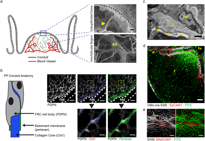
Figure 1. An FRC conduit network facilitates the flow of absorbed lumenal fluids through intestinal PPs.
(a) Scanning electron micrograph of PP following alkali-water maceration to remove cells. top, PP dome region. Conduit (yellow arrow) extending inward from the ECM underlying the dome epithelium (dashed line). bottom, PP interfollicular region. Conduits intersecting a blood vessel (BV) in the PP follicle. Scale bar = 2um. (b) Confocal microscopy of the dome region of a PP stained with anti-PDPN, anti-Collagen Type I, and anti-Perlecan. Follicle associated epithelium marked by dotted lines. Scale bar = 20um. Inset Scale bar = 5um. (c) Transmission electron micrograph depicting the anatomy of a PP conduit. Basement membrane marked by yellow arrows. Scale bar = 0.5um (d) Multiphoton microscopy of a PP from a VillinCre-SSB-BFP mouse after oral gavage with soluble FITC and counterstained with anti-EpCAM1. IF = interfollicular region, F = follicle, Ep = epithelium. Scale bar = 50um (e) Multiphoton microscopy of a PP HEV. The mouse was orally gavaged with soluble FITC and the HEV stained by in vivo labeling with anti-MAdCAM before perfusion fixation. Data representative of images taken from at least 2 (a-c) or 3 (d,e) independent experiments.

