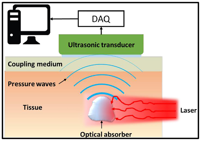
Figure 1.
Basic imaging scheme of PAT. Firstly, laser light is delivered to excite the target (optical absorber) in the tissue. The absorbed optical energy is partially or completely converted into heat, inducing a transient thermal expansion and local pressure rise. The pressure alterations propagate in tissues in the form of ultrasound waves, which are detected by ultrasonic detector(s) placed outside the tissue. After amplification and digitization, the detected signals are used to reconstruct the final PAT image reflecting optical absorption contrast inside tissues.

