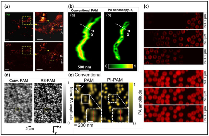
Figure 2.
Photoacoustic microscopy with sub-diffraction resolutions. (a) Melanin distribution imaged by two-photon PAM (2PA) (top) and one-photon PAM (1PA) (bottom) (1). (b) Mitochondria imaged by PA nanoscopy based on optical absorption saturation (2). (c) Red blood cells imaged by Grüneisen-relaxation-based PAM (GR-PAM) (top) at three different depths (3). (d) Reversibly-switchable PAM (RS-PAM) (right) has superior resolution on bacteria (left) (6). (e) Nanoparticles imaged by photobleaching-based PAM (PI-PAM) (right) (7).

