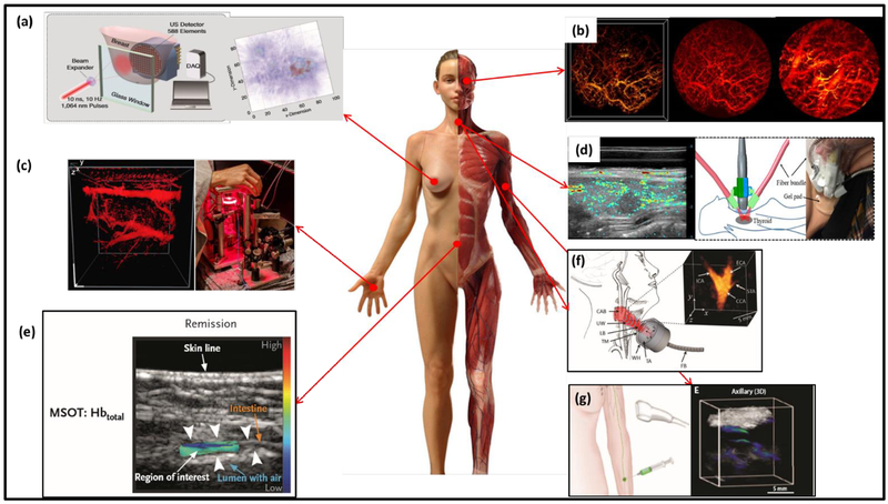
Figure 5.
Representative PAT applications on humans. (a) Twente photoacoustic mammoscopy for breast cancer imaging; left: overview of the system, right: MAP image of the breast showing the lesion clearly on the right side (233). (b) In vivo imaging of oral vasculature; left: 3D display of vessels in a human lip, middle: corresponding MAP image, and right: PA image of the deeper region (264). (c) In vivo imaging of the palm vessels; left: 3D image of the vascular network; right: the imaging system schematics (263). (d) Dual-mode PA/ultrasound imaging of human thyroid. Left: right lobe papillary thyroid cancer image. Right: schematics of the imaging system (265). (e) Illustration of the MSOT of inflammatory bowel disease (267). (f) vMSOT of the human carotid artery, showing the carotid bifurcation (266). (g) MSOT of SLN using ICG as the exogenous contrast (268).

