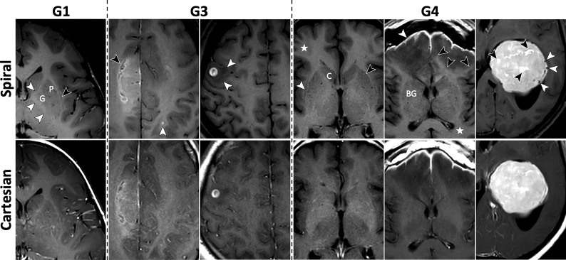
FIG 2.
Images for metrics: subjective SNR (M2), GM/WM contrast (M3), and image sharpness (M4). See On-line Fig 2 for corresponding histograms of scores. Column 1, Spiral has higher SNR and GM/WM contrast as demonstrated by the sharper demarcation of the putamen (P) and globus pallidus (G) from surrounding WM tracts. The internal capsule, claustrum, external capsule, and extreme capsule are clearly distinguishable on spiral (white and black arrows), with sharper margins and higher contrast compared with Cartesian. The cortex and subcortical WM show higher contrast as well. Column 2, Grade II isocitrate dehydrogenase (IDH) mutant astrocytoma following radiation therapy is seen in the right hemisphere, showing a more detailed appearance on the spiral (black arrow). The higher spiral SNR also enables confident detection of an enhancing metastasis in the left parietal lobe (white arrow); this lesion is only faintly visible on the Cartesian. Column 3, Spiral shows better defined borders of the vasogenic edema (white arrows) surrounding the contrast-enhancing metastasis. The overall appearance of the lesion is the same on the 2 sequences. Column 4, Spiral demonstrates higher GM/WM contrast, particularly conspicuous by comparing details of the basal ganglia and delineation of the caudate head (C), lateral borders of the anterior putamen (black arrow), and claustrum (white arrow). The cerebral cortex and subcortical WM are better distinguished (star). Column 5, Meningitis (white arrow) and early cerebritis. The extent of the vasogenic edema is easier to assess on spiral due to the higher contrast and sharper boundary with the WM (black arrows). The contrast between WM and deep GM structures (BG) and the cortex (star) is again higher with spiral. Column 6, Spiral GM/WM contrast is generally higher, while Cartesian has less distinguishable GM/WM boundaries. Internal structure of the intraventricular meningioma shows more details, evident by smaller areas of increased enhancement (black arrows) and vascular structures (white arrows) due to spiral flow compensation.

