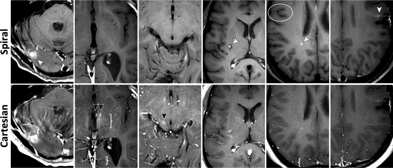
FIG 3.
Images for metrics: individual lesion conspicuity (M5), diagnostic preference for detecting abnormal enhancement (M6), and overall intracranial IQ (M7). See On-line Fig 3 for corresponding histograms of scores. All images are taken from G1. Column 1, Enhancing lesion (white arrow) and surrounding area are better evaluated due to spiral flow artifact reduction. Columns 2 and 3, Misleading hyperintense vascular artifacts (black arrows) are removed in spiral, along with flow-ringing artifacts (braces). Column 4, Cavernous hemangioma in the right basal ganglia (white arrow). The peripheral vascular component is well-separated from the central, contrast-enhancing part by spiral flow suppression. Column 5, An enhancing demyelinating lesion (white arrow) is better depicted on spiral. The spiral was acquired before the Cartesian in this case; thus, the difference is not a result of delayed enhancement. Better delineation of the cortex and higher GM/WM contrast are seen again (circle), along with removal of flow ringing (brace). Column 6, Postsurgical residual enhancement (white arrow) has higher signal and better delineation in spiral, which increases diagnostic confidence.

