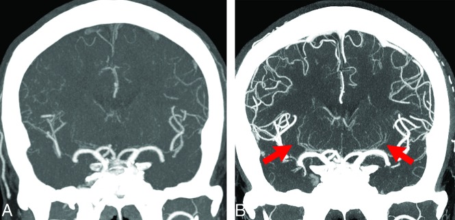
FIG 2.
A 49-year-old man with anterior cerebral artery dissection. A, C-CTA of a coronal partial MIP (20 mm) image. B, UHR-CTA of a coronal partial MIP (20 mm) image. Bilateral LSAs, particularly left LSAs, are not depicted at all on C-CTA; however, they are depicted from the proximal-to-distal side in UHR-CTA (arrows). Peripheral branches of the anterior cerebral artery, MCA, and posterior cerebral artery in UHR-CTA are also observed more clearly compared with C-CTA.

