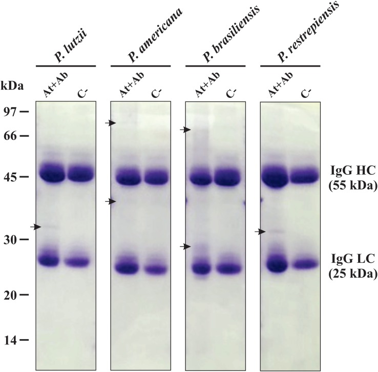
FIGURE 2.
Electrophoretic profile of affinity chromatography. One-dimensional (1-D-SDS-PAGE) gel of proteins eluted from Sepharose beads-IgG. Immunoprecipitation using serum from immunized mice (At + Ab) and negative control (C−). Top row: Isolated representatives of Paracoccidioides species (P. lutzii, P. americana, P. restrepiensis, and P. brasiliensis). The arrows indicate the presence of exoantigens after purification when compared to the negative control. At, antigen; Ab, antibody; IgG HC, immunoglobulin G heavy chain; IgG LC, immunoglobulin G light chain. Electrophoresis stained with Coomassie blue.

