
Figure 4.
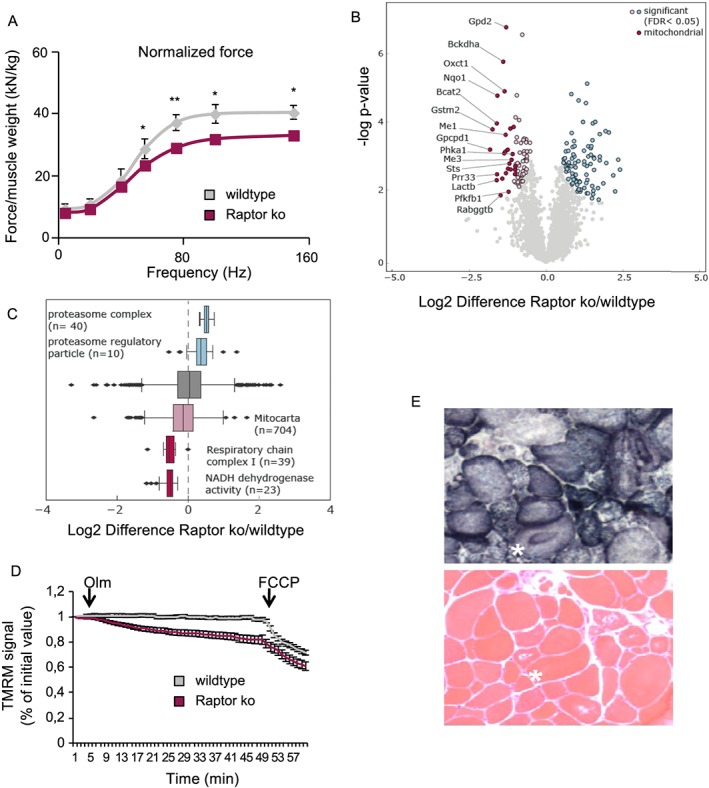
Long‐term deletion of Raptor leads to mitochondrial dysfunction with reduced force production. (A) Normalized force measured in vivo is significantly reduced in Raptor k.o. mice (n = 6 per group). (B) Volcano plot of differentially regulated proteins in Raptor k.o. muscles 7 months after the deletion of Raptor (n = 3). Significantly regulated proteins are marked in blue and light red (FDR < 0.05, s0 = 0.1, number of permutations: 500). Mitochondrial proteins are highlighted in dark red. (C) Box plot of log2 Raptor k.o./wild‐type ratios of proteins associated with the proteasome, the respiratory chain complexes or the mitochondrion. (D) Mitochondrial dysfunction revealed by TMRM. Oligomycin (Olm) and the protonophore FCCP were added at the indicated timepoints (n = 30/group). (E) Mitochondrial morphology is drastically altered in Raptor k.o. muscles. Data are shown as mean ± SEM. Statistical analysis was performed using two‐tailed Student's t‐test. Statistical significance: *P < 0.05 and **P < 0.01.