
Figure 7.
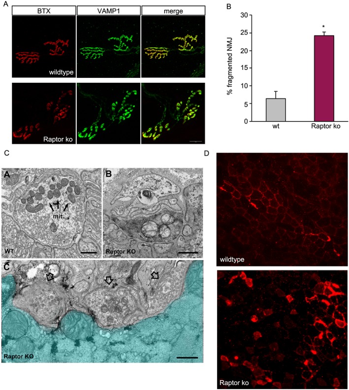
Reduced mTORC1 signalling leads to neuromuscular junction fragmentation. (A) NMJs in wt muscles show their normal pretzel‐like shape. Raptor k.o. mice show numerous NMJs in which both presynaptic (VAMP1) and postsynaptic [acetylcholine receptor (BTX)] structures accumulate in individual, non‐connected aggregates. (B) Quantification of the percentage of fragmented NMJs in wt and long‐term Raptor k.o. mice (n = 4 mice/group). (C) Electron microscope images of NMJs in wild‐type and Raptor k.o. muscles. Note the altered presynaptic structures in Raptor k.o. muscles. (D) Staining for acetylcholinesterase in wild‐type and Raptor k.o. muscles. Data are shown as mean ± SEM. Statistical analysis was performed using two‐tailed Student's t‐test. Statistical significance: *P < 0.05.