
Figure 8.
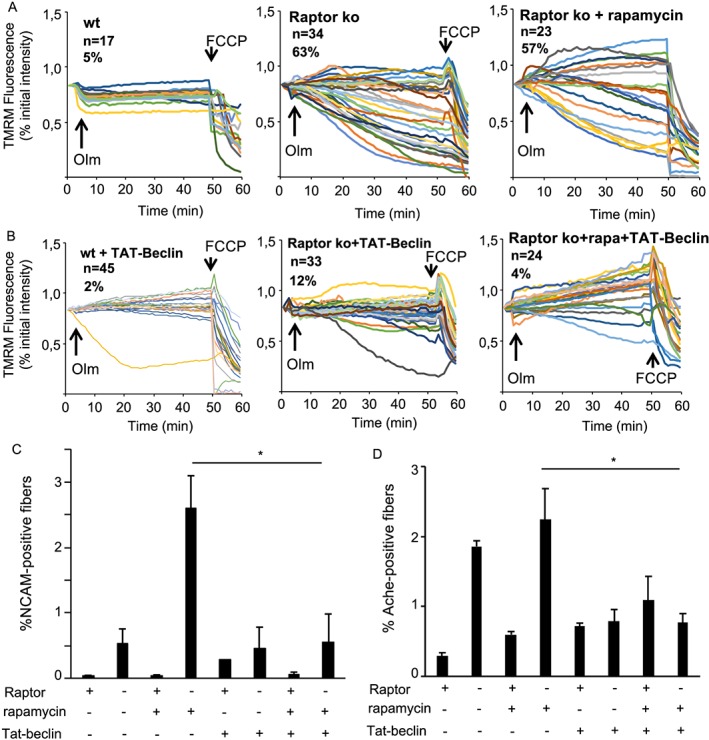
Reactivation of autophagy by Tat‐beclin1 prevents the appearance of NCAM‐positive fibres in Raptor k.o. muscles. (A) TMRM measurement of mitochondrial membrane potential in isolated fibres from the FDB muscle. Raptor k.o. fibres, both with and without rapamycin treatment, show a significant mitochondrial depolarization after oligomycin addition. (B) Mice treated for 2 weeks daily with Tat‐beclin1 completely prevented the mitochondrial dysfunction in Raptor k.o. fibres. (C, D) Gastrocnemius muscles taken from mice treated for 2 weeks with rapamycin and Tat‐beclin1 and stained for NCAM or AchE. No increase in NCAM or AchE staining in Raptor k.o. mice after co‐treatment with rapamycin and Tat‐beclin1 (n = 4–6 muscles/group). Data are shown as mean ± SEM. Statistical analysis was performed using two‐tailed Student's t‐test and two‐way ANOVA when required. Statistical significance: *P < 0.05.