
Figure 3.
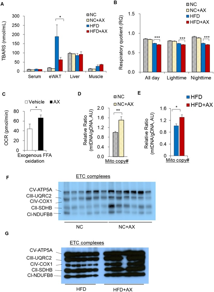
AX potentially enhanced the mitochondrial number and fatty acid utilization in skeletal muscle in HFD obese C57BL/6J mice. (A) AX partially reduced the oxidative stress in the adipose tissue of HFD obese C57BL/6J mice. Oxidative stress markers in the serum, skeletal muscle, eWAT, and the liver of AX‐treated HFD and NC mice compared with non‐AX‐treated HFD and NC mice. Oxidative stress was evaluated based on the production of malondialdehyde and TBARS (n = 3 in each group). (B) The respiratory quotient (RQ) in the mice fed NC, NC+AX, HFD, or HFD+AX for 8 weeks (n = 6 per group). (C) Exogenous free fatty acid (FFA) oxidation rates were measured in C2C12 cells treated with vehicle or 5 μM AX for 24 h, using oxygen consumption rate (OCR) as a readout with/without 40μM etomoxir and/or BSA‐Palmitate (n = 6 per group) using the XFe96 Extracellular Flux. (D,E) Relative mtDNA copy number in the gastrocnemius muscles of the C57BL/6J mice fed NC, NC+AX (D), HFD, or HFD+AX (E) (for 24 weeks (n = 6 per group). (F,G) Western blot analysis of mitochondrial electronic chain transport complexes (ETCs) in the C57BL/6J mice fed NC, NC+AX (F), HFD or HFD+AX (G) for 24 weeks (n = 6 per group). All values are presented as means ± S.E.M. *p < 0.05, **p < 0.01, ***p < 0.001 (HFD vs. HFD+AX) or (NC vs. NC+AX). Statistical analysis was performed using Student's t‐test (A‐E).