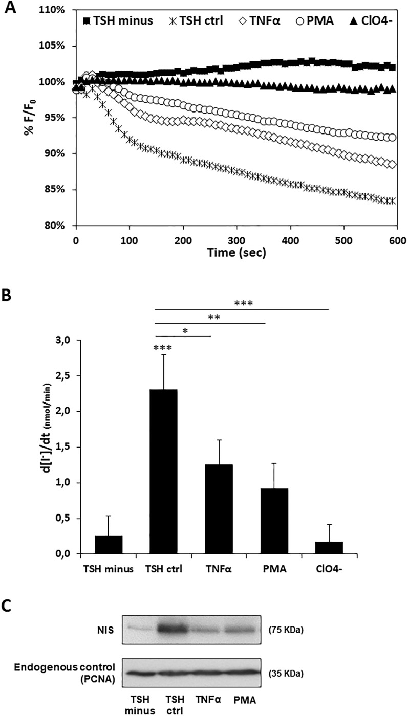
Fig 3. Effect of TNF-α and PMA on TSH-induced iodide uptake and NIS protein levels in Y-PCCL3 cells.
Cells PCCL3 cells stably expressing the YFP-halide sensor were subjected to a 24h starvation period and then treated with TSH for 96h (TSH ctrl), in the presence or absence of either TNF-α or PMA Additional iodide influx assays were performed in the presence of both (24h treatment), and subjected to iodide influx assays and Western blot. TSH and ClO4- (1 mM, 10 min), a competitive inhibitor of iodide uptake by NIS. Representative HS-YFP fluorescence decay traces (A) recorded continuously for 600 seconds, acquiring an image every 10 s, after exposure to 1mM NaI (as described in [30]). Fluorescence (F) was plotted over time as percentage of fluorescence at time 0 (F0). Iodide influx rates (B) calculated by fitting the curves to the exponential decay function to derive the maximal slope that corresponds to initial influx of I− into the cells. Endogenous NIS protein expression upon TSH, TNF-α or PMA treatment was monitored Western Blot using anti-NIS primary antibody. Endogenous PCNA expression was used as loading control. Data are means ± SEM of three independent assays. Comparisons were made using one-tailed Student’s t-tests (*p≤0.05; **p ≤0.01; ***p ≤0.001).

