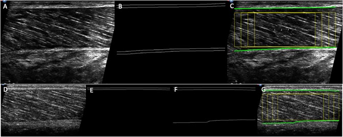
Fig 2.
Examples of raw images of the ultrasound field of view, aponeuroses edge detection and overlaid ROIs (yellow) on a suitable scan (A-C), and on a scan where the lower aponeurosis is imaged with insufficient contrast (D-G). In C and G, the paths of the superficial and deep aponeuroses are also indicated as green lines. When the aponeurosis is imaged with insufficient contrast it is not detected (E). In this case using a higher Tubeness sigma may help detection (F-G), although too high values may cause a slight distortion of the aponeurosis edge.

