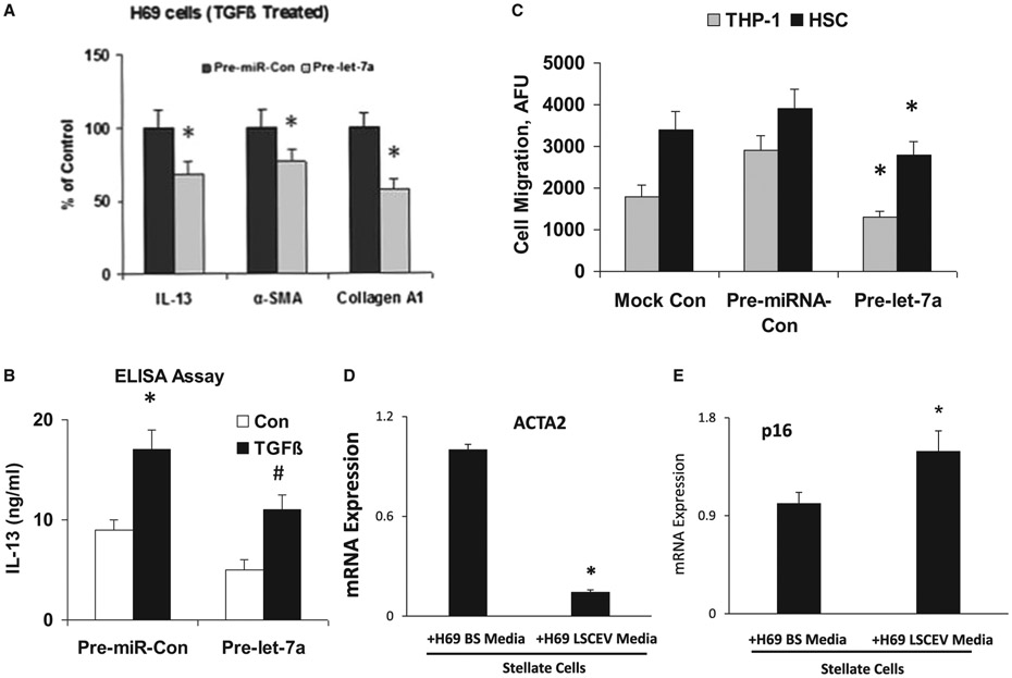
FIG. 7.
Let-7/LSCEV-mediated anti-inflammatory and antifibrotic interactions between cholangiocyte and human macrophages/human hepatic stellate cells. (A,B) Pre-let-7a treatment inhibits TGF-β-stimulated cholangiocytes and suppresses the migration of THP-1 cells. (A) Effects of Pre-let-7a on the mRNA expression of inflammation/fibrosis markers in H69 human cholangiocytes. H69 cells were transfected with 100 nM Pre-miRNA-Con or 100 nM Pre-let-7a. After incubation in DMEM with 1% FCS, the cells were treated with TGF-β (25 mM) for 48 hours. Cellular RNAs were isolated, and the expressions of IL-13, α-SMA, and Col1A1 mRNA levels were examined by real-time quantitative PCR. GAPDH mRNA was used for normalization. (B) Effects of Pre-let-7a on the expression of cytokine IL-13 at the protein level. Conditioned medium was collected from TGF-β-stimulated H69 cells (25 mM for 48 hours), and enzyme-linked immunosorbent assay was performed to assess IL-13 expression. (C) Migrations of the human monocyte cell line THP-1 and human HSCs were suppressed by conditioned media from Pre-let-7a-transfected H69 cells. A total of 5 × 10⁵ THP-1 cells or 5 × 10³ human HSCs were placed in the upper chamber, and conditioned medium from TGF-β-stimulated H69 cells transfected with Pre-miR-Con or Pre-let-7a was added to the lower chamber. After 3 hours of incubation, THP-1 cells or HSCs that migrated toward the lower chamber were detected by fluorescent dye. Relative fluorescence units (AFUs) are indicated (versus 1% FCS). All results are shown as mean ± SEM. A minimum of three replicates were performed for each set of experiments to compile the data as presented. *P < 0.05 relative to Pre-miR-Con group. To evaluate the interactions among LSCs, cholangiocytes and HSCs, H69 human cholangiocytes were incubated with LSCEVs for 48 hours; the media was then changed to new media for 24 hours. The H69 media was removed and used to treat stellate cells for 48 hours. The stellate cells were then used to extract mRNA and perform real-time quantitative PCR for the fibrosis marker, ACTA2 (D), and the senescence marker, p16 (E). Abbreviations: ELISA, enzyme-linked immunosorbent assay; GAPDH, glyceraldehyde 3-phosphate dehydrogenase.

