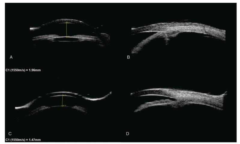
Figure 1.

A and B, UBM showed shallowing of the AC and angle closure in the right eye. C and D, UBM showed shallowing of the AC and angle open in the left eye. B and D showed inferior anterior chamber angle. AC = anterior chamber, UBM = ultrasonographic biomicroscopy.
