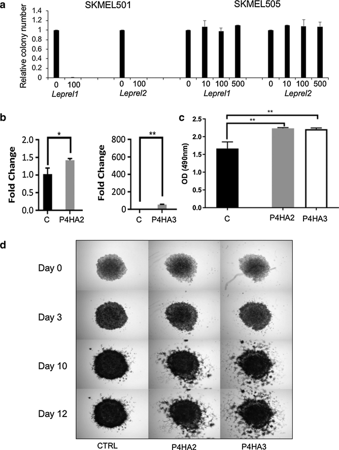
Figure 3. Collagen prolyl 3-hydroxylase and collagen prolyl 4-hydroxylase have opposing effects on melanoma progression.
(a) Ectopic expression of Leprel1 and Leprel2 in cells lacking endogenous expression blocks proliferation. (b) Quantitative PCR analysis in PMWK engineered to express of P4HA2 and P4HA3. Data are mean ± standard error of the mean (n = 3). Significance was tested using one-way analysis of variance with Dunnett’s post-hoc testing.(c) P4HA2 and P4HA3 overexpression significantly increase proliferation in PMWK cells. Graphs shows OD at 490 nm 6 days post treatment. Data are mean ± standard error of the mean over biological repeats (n = 2) performed in triplicate. Significance was tested using two-way analysis of variance with Tukey’s post-hoc testing. (d) P4HA2 and P4HA3 overexpression increase the invasiveness relative to control (CTRL) of PMWK early radial growth phase melanoma cells. Images are representative of two independent experiments performed in triplicate. OD, optical density.

