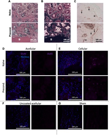
Fig. 6. Human ECs and MSCs engraft into new bone tissue.

(A to C) Histological sections stained with (A) H&E, (B) Masson’s trichrome, and (C) anti-human CD31. B and O denote new bone and osteoid, respectively, and black arrows denote vasculature of human origin. Scale bars, 100 μm. (D to G) Immunofluorescence staining of histological sections for anti-human DLX5 (magenta) and cell nucleus (blue). (D and E) (Top) Naïve and (bottom) preconditioned (D) acellular and (E) BMA-loaded scaffolds compared to (F) uncoated acellular and (G) sham controls. Scale bars, 100 μm.
