
Fig. 3. MD simulation of the undocked INX-6 hemichannels embedded in phospholipids and unassigned densities in cryo-EM maps.
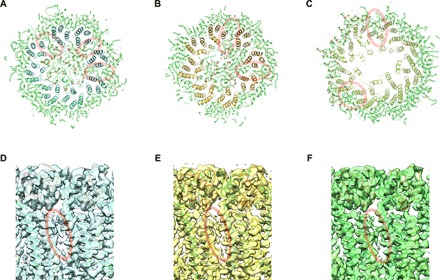
(A to C) The final models of the three atomic structures obtained by cryo-EM in this work after independently performed MD simulations in POPC for 100 ns [120 ns for (B)]. Slab sections of WT INX-6 in a nanodisc (A), INX-6ΔN in a nanodisc (B), and WT INX-6 in detergent (C) corresponding to the transmembrane domain along with POPC molecules are viewed from the cytoplasmic side. POPC models that are inserted in the space between adjacent subunits are indicated by red circles. (D to F) Unassigned densities (red circles) observed in the space between transmembrane helix bundles of adjacent subunits of WT INX-6 in a nanodisc (D), INX-6ΔN in a nanodisc (E), and WT INX-6 in detergent (F) viewed from the orientation horizontal to the membrane plane.