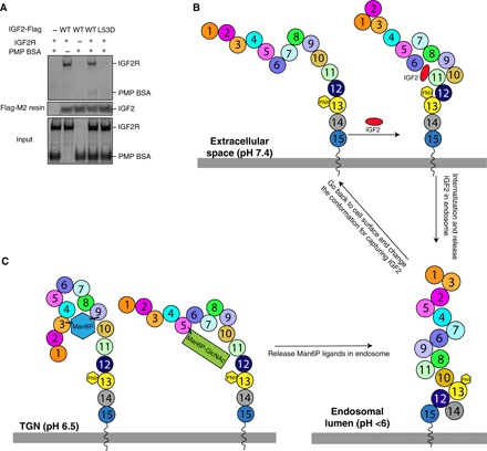
Fig. 5. The binding sites of IGF2R to Man6P and IGF2 are independent.

(A) Pull-down assay between IGF2, PMP-labeled BSA, and IGF2R. IGF2 is conjugated onto Flag-M2 resin. The proteins are shown on SDS-PAGE with Coomassie blue staining. (B) Working model of IGF2R for IGF2 recognition, postulating a molecular mechanism of how IGF2R changes its conformation to recognize IGF2 by using its domains at different pH values. (C) A speculation of how Man6P substrates are recognized by IGF2R. TGN, trans-Golgi network.
