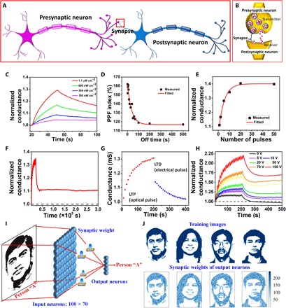
Fig. 5. Photonic synapse performance and facial recognition.

(A) Anatomy of two interconnected human neurons via a synapse (red box). (B) Schematic representation of biological synapses. (C) Transient characteristic of the device (VD = 0.5 V and VG = 10 V) showing the change in conductance due to a single pulse of light of pulse width 30 s for varying light intensity. (D) PPF index of the device (VD = 0.5 V and VG = 10 V) due to varying off time between two consecutive light pulses having on time of 5 s. (E) Transient characteristic of the device (VD = 0.5 V and VG = 10 V) showing the change in conductance due to varying number of light pulses having on and off time of 5 and 5 s, respectively. (F) Retention of the long-term potentiated device (VD = 0.5 V and VG = 10 V) for 3 × 10³ s after application of 20 optical pulses (on and off time of 5 and 5 s, respectively). (G) Nonvolatile synaptic plasticity of the device (VG = 10 V) showing LTP by train of optical pulses (on and off time of 5 and 5 s, respectively) at VD = 0.5 V and LTD by a train of electrical pulses (−0.5 V, on and off time of 1 and 1 s, respectively) at VD. (H) Gate-dependent transient characteristic of the device (VD = 0.5 V) after application of 20 optical pulses (on and off time of 5 and 5 s, respectively).(I), Neuron network structure for face recognition. Photo credit: Sreekanth Varma and Basudev Pradhan, UCF. (J) Real images (top) for training and the synaptic weights of certain corresponding output neurons (bottom). Photo credit (from left to right): Sreekanth Varma and Basudev Pradhan, UCF; Avra Kundu and Basudev Pradhan, UCF; Basudev Pradhan, UCF; and Basudev Pradhan, UCF.
