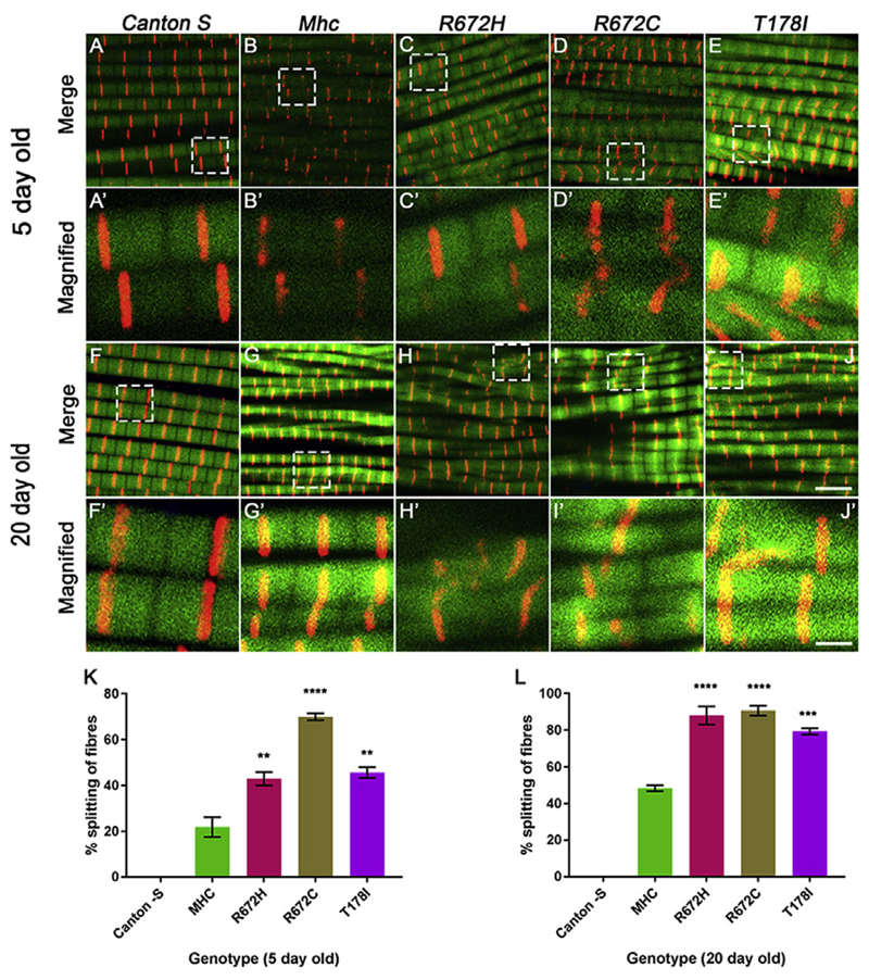
Fig. 5. Expression of FSS mutant Mhc transgenes lead to increased fiber splitting 5 and 20 days post-eclosion.
Representative dorsal longitudinal muscles (DLMs) from 5 to 20 days post-eclosion wild type Canton S (A-A′, F-F′), Mhc1/CyO; Mef2GAL4/UAS-Mhcwt (B-B′, G-G′), Mhc1/CyO; Mef2GAL4/UAS-MhcR672H (C-C′, H-H′), Mhc1/CyO; Mef2GAL4/UAS-MhcR672C (D-D′, I-I′) and Mhc1/CyO; Mef2GAL4/UAS-MhcT178I (E-E′, J-J′), labeled by immunofluorescence for Kettin marking the Z-disc (red), and Actin labeling the thin filament (green). A-E′ are DLMs from 5 days post-eclosion flies and F-J′ are DLMs from 20 days post-eclosion flies. The boxed regions in the images A-E and F-J are magnified and shown in A′-E′ and F′-J′ respectively. Quantification of the percentage of split fibers as a proportion of total fibers for DLMs from each genotype at 5 days (K) and 20 days (L) post-eclosion are shown. The graphical data is presented as mean ± standard error of the mean using a minimum of 5 replicates. Scale bar in J is 5 μm and J′ is 1 μm.

