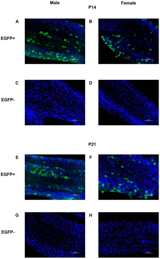
Figure 2. Enhanced green fluorescent protein (EGFP) in the trigeminal ganglion of postnatal day (P) 14 and P21 male and female OXTR:EGFP transgenic mice:
Fluorescent microscopy for EGFP (green) and DAPI (blue) in OXTR:EGFP+ (A, B, E, F,) and OXTR:EGFP−(C, D, G, H) trigeminal ganglion at P14 (A-D) and P21 (E-H) males (A, C, E, G) and females (B, D, F, H). EGFP is present in the trigeminal ganglion of OXTR:EGFP+, but not OXTR:EGFP−, P14 and P21 males and females. Images taken at 20X magnification. Scale bar = 100μm.

