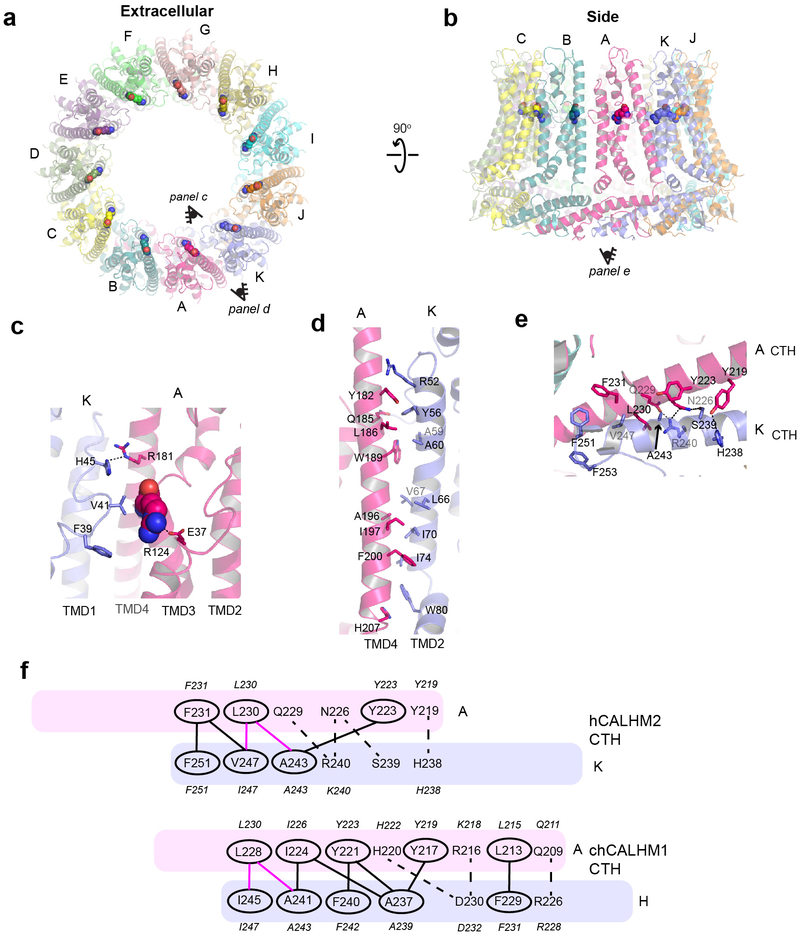
Extended Data Fig. 7. Interaction of hCALHM2 subunits.
a-b, The hCALHM2 structure viewed from the top of extracellular region (a) and the side of the membrane (b). Shown in spheres are the Arg124 residues at the equivalent position to chCALHM1 Asp120 or hCALHM1 Asp121. c, Arg124 (sphere) and surrounding residues (sticks) form polar and hydrophobic interactions to mediate inter-subunit interactions. d-e, The inter-subunit interactions between TMD2 and TMD4 (d) and CTHs (e). f, The schematic presentation of the interactions between two CTHs (magenta and slate blue) in hCALHM2 (top) and chCALHM1 (bottom). Polar and van der Waals interactions mediated by hydrophobic residues (ovals) are shown as dashed and solid lines, respectively. The lines in magenta are the conserved interactions between chCALHM1 and hCALHM2. The residues in italic are the equivalent ones in hCALHM1.

