
Figure 6.
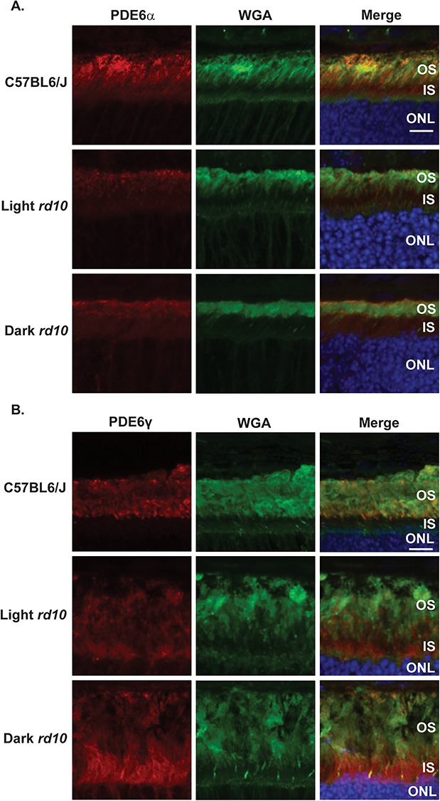
PDE6γ but not PDE6α is mislocalized in both light and dark reared rd10 mice. (A) Immunofluorescence microscopy images of retinal cross sections from the light and dark reared rd10 mice along with a C57BL6/J wild-type control after probing with antibody directed against PDE6α (red), WGA (rod photoreceptor OS marker shown in green) and DAPI (blue) nuclear counterstain. Scale bar = 10 μm. (B) Immunofluorescence microscopy images of retinal cross sections from the light and dark reared rd10 mice along with a C57BL6/J wild-type control after probing with antibody against PDE6γ (red), WGA (green) and DAPI (blue). Scale bar = 10 μm.