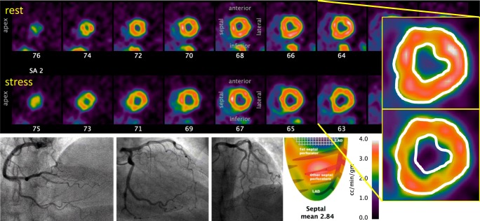
Fig. 2.
Subendocardial angina with non-obstructive epicardial arteries. This 68-year-old woman suffered from exertional angina and dyspnea for several years, prompting an invasive angiogram that demonstrated at most mild atherosclerosis. Echocardiography demonstrated normal left ventricular (LV) function and LV hypertrophy but no significant valvular disease. Due to persistent symptoms, she underwent a cardiac PET scan with dipyridamole stress during which she developed severe angina. An excellent average stress flow of over 2.8 cc/min/g excludes microvascular dysfunction. While no regional defect is present, the inset images copy the resting epicardial and endocardial borders onto the stress tomographic images. A nearly circumferential subendocardial perfusion defect explains her symptoms and would not have been identified by epicardial assessment of FFR, flow reserve, or myocardial resistance

