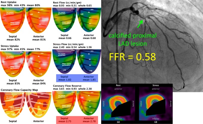
Fig. 3.
Absolute versus relative flow. This 78-year-old man had mild angina from a culprit lesion in the left circumflex, not shown since the focus is on the physiology of the left anterior descending (LAD) artery. The anterior and septal quadrants show no relative defect, either for peak uptake or transmural perfusion. Average hyperemic flow exceeded 1.8 cc/min/g, far above the thresholds for myocardial ischemia. Coronary flow reserve was over 2.7, excluding microvascular disease. Wall motion in these quadrants was normal and serum caffeine was negative. Despite intact flow to the LAD distribution, invasive angiography showed a severe, calcified lesion in the proximal vessel with an invasive FFR of 0.58. After revascularization for the angina culprit in another vessel, including a mammary artery bypass of the LAD, repeat cardiac PET showed an increase in flow to 2.8 cc/min/g, indicating a 1.8/2.8 = 0.64 relative reduction in flow similar to the pre-revascularization FFR prediction. In this case, relative flow indeed decreased by approximately 40% due to this lesion, but started from such a high level that its reduction did not produce ischemia, only a pressure gradient

