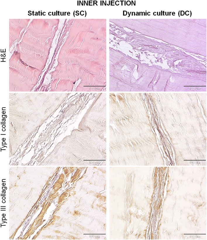
Figure 6.
H&E and collagen type I and III immunostaining of samples, 7 days after static or dynamic culture. Within the cell injected fibers, the newly synthesized extracellular matrix appeared thinner, looser and less organized in the SC constructs, whereas in the DC group it was more dense and structured. The matrix deposited by DC was qualitatively little more positive for type I collagen than that synthesized by SC, while type III collagen was comparable between the two groups. Magnification ×200, scale bars 100 µm.

