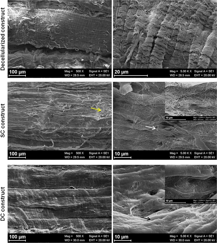
Figure 8.
SEM imaging. At the lowest magnification (100 µm), decellularized and SC tendons showed the classical tendon morphology with orderly, parallel collagen fiber bundles, even if some of them emerged from the matrix not following the general organization (yellow arrow). At higher magnification (20 µm), cells were clearly visible on the surface of the fibrillar matrix of the SC constructs with protrusions departing from rounded cell bodies (white arrow). Whereas, in the DC constructs, cells appeared completely embedded within the dense matrix and cells were not clearly recognizable (black arrow).

