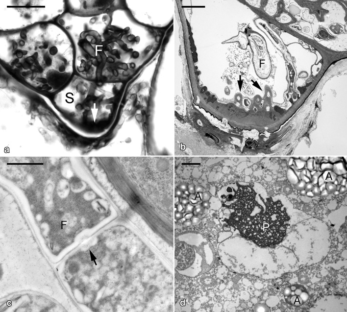
Fig. 2.
Micrographs showing the germinating embryo of G. elata associated with Mycena. a Light micrograph of the suspensor end cell (S) colonized by fungal hyphae (F) with cell wall thickening (arrow).Scale bar = 10 μm. b Ultrastructural view of the suspensor end cell showing the papillae-like cell wall thickening (arrows) corresponding to the entry of fungal hyphae. Scale bar = 2 μm. c At this stage, the intact fungal hypha (F) is present in the primarily colonized cells. The dolipore septum (arrow) can be observed at the junction between fungal cells. Scale bar = 1 μm. d In the uncolonized embryo cells, the storage protein bodies (P) are degrading and amyloplasts (A) start to appear. Scale bar = 4 μm

