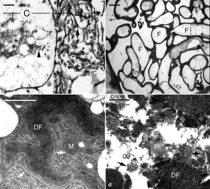
Fig. 4.
Micrographs showing the elongated protocorm of G. elata associated with Mycena. a Light micrograph showing the colonized region of the elongated protocorm. The epidermal cell (E) contains old fungal hyphae (arrow), and fragments of digested fungal hyphae (arrowhead) are visible in the cortical cell (C). Scale bar = 10 μm. b In the epidermal cell of the elongated protocorm, the cytoplasm of old fungal hyphae (F) has degenerated. Scale bar = 2 μm. c In the cortical cell of the elongated protocorm, the digested fungal hyphae (DF) are surrounded by rough endoplasmic reticulum (arrows) and a few mitochondria (M). Scale bar = 1 μm. d The digested fungal hyphae (DF) become fragmented and associated with clusters of vesicles (arrows). Scale bar = 1 μm

