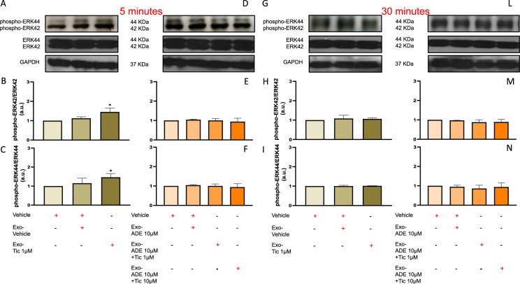
Figure 6.
Long-term pre-treatment for 48 h of normoxic HL1 cardiomyocytes with exosomes (100 μg) derived from hCPCs treated with low-dose ticagrelor (Tic 1 μM) increase levels of phosphorylated ERK42/total ERK42 (B; 42KDa MW) and phosphorylated ERK44/ total ERK44 (C; 44KDa MW) ratios after 5 min. Representative images of cropped densitometric bands of phosphorylated and total ERK 42/44, and glyceraldehyde 3-phosphate dehydrogenase (GAPDH; 37KDa MW) are showed in panel A and D. All full-length blots/gels are presented in Supplemental Fig. 8 panel A. As shown in panels E,F, exosomes released from hCPCs treated in the presence of adenosine (ADE, 10 μM) and EHNA (10 μM) do not induce rising of intracellular phosphorylated ERK42/44 levels. Representative images of cropped densitometric bands of phosphorylated and total ERK 42/44, and GAPDH are showed in panel D. Intracellular levels of phosphorylation of ERK42/44 are normal in HL1 cells after 30 min of treatment with ticagrelor-induced exosomes in the absence (H,I) or presence (L,M) of adenosine (ADE 10 μM) + EHNA (10 μM). Representative images of cropped densitometric bands of phosphorylated and total ERK 42/44, and GAPDH are showed in panel G and L. All full-length blots/gels are presented in Supplemental Fig. 8 panel B. Levels of ERK42/44 are normalized on GAPDH levels and ratios are expressed as arbitrary units (a.u.). All measurements are mean ± SD. *p < 0.05 vs. untreated condition (Vehicle: sterile phosphate buffer solution).

