Figure 2.

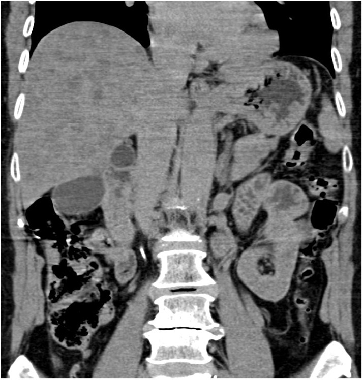
Abdominal computed tomography, non-enhanced coronary scan. Markedly inhomogenous liver with map-like hyperdense areas and multiple hypodense focal changes.

Abdominal computed tomography, non-enhanced coronary scan. Markedly inhomogenous liver with map-like hyperdense areas and multiple hypodense focal changes.