
Figure 2.
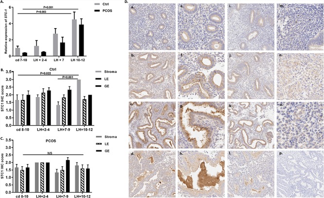
Expression of STC-1 in human endometrium. (A) Quantitative reverse-transcription polymerase chain reaction analysis of endometrial samples from healthy control women (gray bar) and women with PCOS (black bar) collected at cd 7–10 (nctrl = 7, nPCOS = 7), LH + 2–4 (nctrl = 5, nPCOS = 5), LH + 7 (nctrl = 7, nPCOS = 7), and LH + 10–12 (nctrl = 5, nPCOS = 7) revealed a significant increase in endometrial STC-1 gene expression from the PE (cd 7–10) toward the LSE (LH + 10–12) in both control samples (P = 0.003) and samples from women with PCOS (P = 0.001). Data are presented as mean ± standard error of the mean (SEM). (B) Semi-quantitative immunohistochemistry data on STC-1 protein expression in different endometrial cell compartments (stromal cells (stroma), luminal epithelium (LE), and GE) from control samples assessed at cd 8–10 (n = 6), LH + 2–4 (n = 7), LH + 7–9 (n = 6), and LH + 10–12 (n = 7) revealed the highest expression in the LSE (LH + 10–12) compared with the PE (cd 8–10; P = 0.022) and MSE (LH + 7–9; P = 0.001) in stromal cells, whereas (C) there was no significant difference between the cycle phases in cells from women with PCOS. (D) STC-1 expression in different cycle phases studied by immunohistochemistry in control women (a–h) and in women with PCOS (i–l). (a) PE (cd 8–10); (b) ESE (LH + 2–4); (c) MSE (LH + 7–9); (d) LSE (LH + 10–12). Higher magnification images from control women (40×) of GE shows that in PE, STC-1 is localized to the GE cells (e), and in ESE it is concentrated in the apical parts (f) from which it is gradually secreted into the lumen during the MSE (g) and LSE (h). The stromal STC-1 staining was weak in PE (m) and significantly stronger in LSE (n), but in PCOS, the staining intensity was similar across the cycle phases. (o) LSE stroma of a PCOS case; (p) negative control staining.