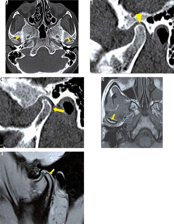
Figure 15.
A-E) Regressive remodelling of condyle. In a patient with left-sided internal derangement, axial (A) and sagittal oblique (B, C) noncontrast computed tomography images show reduced antero-posterior dimension of left condyle (yellow arrow) with maintained vertical height suggestive of regressive remodelling. Right condyle shows normal volume (yellow arrowhead). Right axial T1W (D) and sagittal oblique proton density fat saturation (E) images in another patient with internal derangement show reduced condylar volume (yellow arrow)

