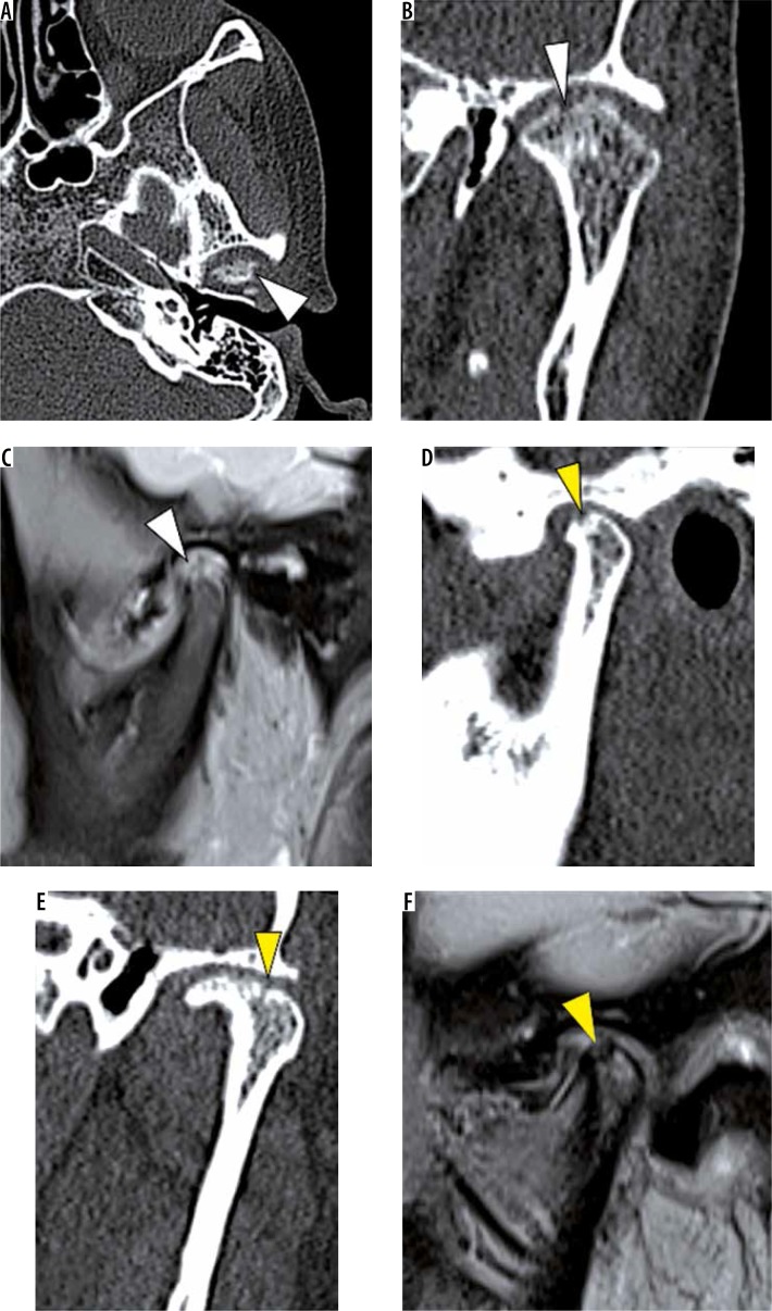
Figure 16.
Erosions. In a patient with active erosive degenerative joint disease (DJD), axial (A) and coronal oblique (B) noncontrast computed tomography (NCCT) and sagittal oblique proton density fat saturation (PD-FS) (C) images show multiple cortical erosions (white arrowheads) with loss of condylar volume. In another patient with incomplete repair phase of DJD, sagittal and coronal oblique NCCT (D, E) and sagittal oblique PD-FS (F) images show loss of condylar volume and shallow concave erosions with a recorticated surface (yellow arrowheads)

