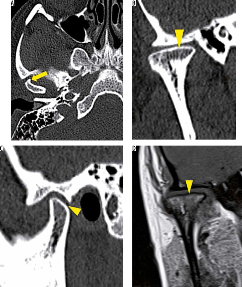
Figure 17.
A-D) Osteophytosis and flattening with loss of height. In a patient with late stage of degenerative joint disease , axial (A), coronal oblique (B), and sagittal oblique (C) and T1W MR (D) images reveal osteophytosis (yellow arrow) and flattening of condyle with loss of vertical height (yellow arrowheads)

