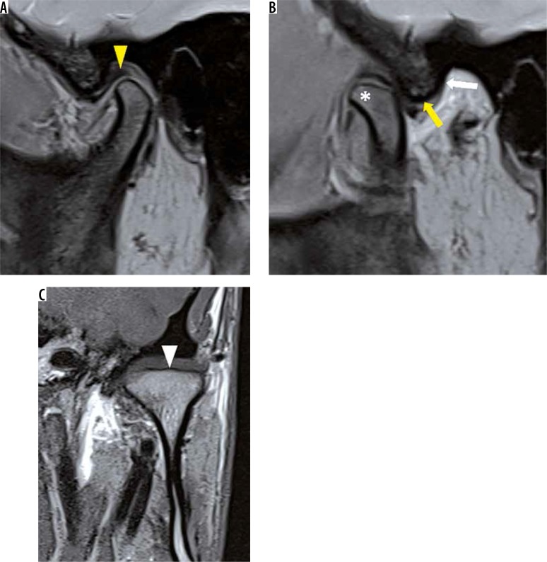
Figure 18.
Hypermobile temporomandibular joint. A) In a 25-year-old patient who presented with multiple episodes of open lock on left side, closed mouth sagittal oblique proton density fat saturation image reveals a normal disc (yellow arrowhead). B) Open mouth image shows increased range of motion where the condyle (asterisk) is seen reaching antero-superiorly to the crest of articular eminence (yellow arrow). Steep (vertical) posterior slope of AE (white arrow) is also noted. C) Coronal oblique T1W image shows flattening of condyle (white arrowhead)

