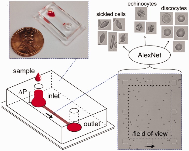
Figure 3.
Automated measurement of RBC morphology. A microfluidic device fabricated to allow RBCs to arrange in a single layer under flow is used to acquired multiple images of sickle RBCs. The images are then classified as sickle RBCs, normal RBCs, or RBCs with other aberrant morphologies (e.g. echinocytes) using a pre-trained convolutional neural network (AlexNet). Arrows indicate flow direction.61 (A color version of this figure is available in the online journal.)

