Figure 1.
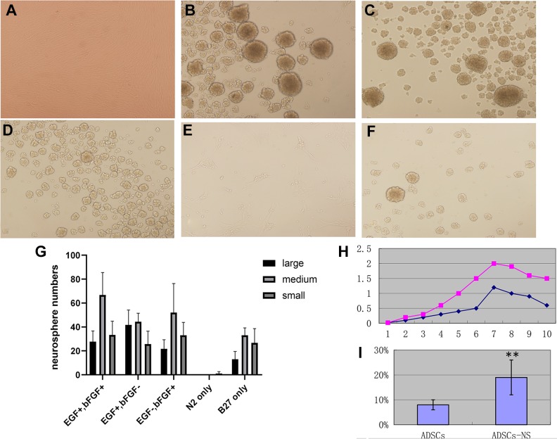
Generation of adipose-derived stem cell (ADSC)-derived neurospheres. (A) ADSCs at passage 3, phase contrast image, 100x. (B)–(F) ADSC-derived neurospheres were generated after 12 hours of induction using different induction conditions. Phase contrast image 100x. (B) ADSC-derived neurospheres induced with epidermal growth factor (EGF) 20 ng/ml, basic fibroblast growth factor (bFGF) 20 ng/ml, and N2 and B27 supplements; (C) EGF+bFGF− regimen with Dulbecco’s modified eagle medium: nutrient mixture F-12 (DMEM/F12), EGF 20 ng/ml, no bFGF, plus N2 and B27 supplements; (D) EGF-bFGF+ regimen with DMEM/F12, bFGF 20 ng/ml, no EGF, plus N2 and B27 supplements; (E) N2 only: DMEM/F12 with N2 supplement only, no EGF or bFGF; (F) B27 only: DMEM/F12 with B27 supplement only, no EGF or bFGF. (G) Statistical analysis of ADSC-derived neurosphere formation assay. Neurospheres were arbitrarily divided into large, medium, and small neurospheres, and scored in six random fields under a microscope. The results represent three independent experiments. (H) Growth curve of ADSC-derived neurospheres in comparison with commercially available human neural stem cells (NouvNeu hNSC, Catalogue No. NC0001, iRegene). The y-axis represents the absorbance value, the x-axis represents days in ex vivo culture. The purple dot plot represents human neural stem cells and the blue dot plot represents ADSC-derived neurospheres. (I) Annexin V and propidium iodide (PI) apoptosis analysis of primary neurosphere formation, in comparison with ADSCs. Apoptosis rate is expressed as mean±SD. ** p<0.01.

