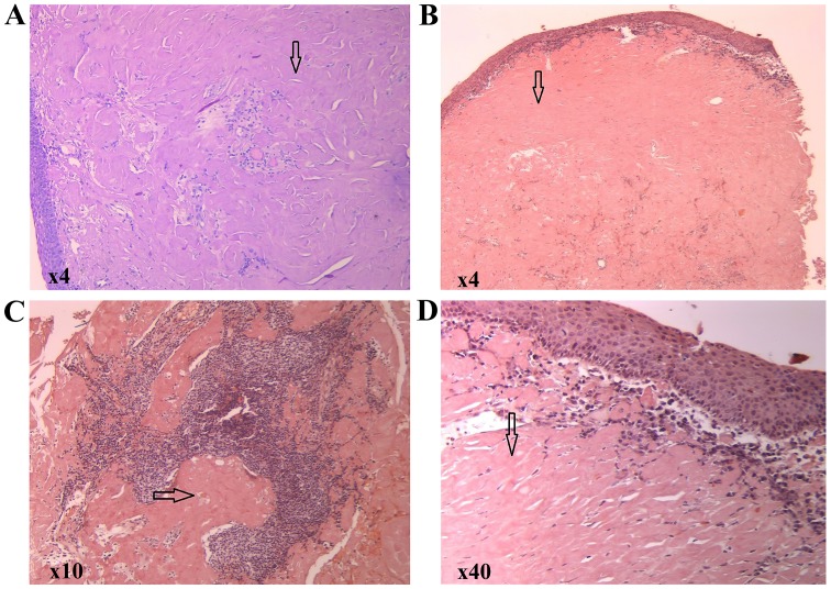
Figure 3.
(A) Hematoxylin and eosin staining showing an eosinophilic amorphous material in the connective tissue beneath the intact epithelium (magnification, x4). (B-D) Congo red staining showing a red homogenous material (arrows) under light microscopy (magnification, x4, x10 and x40, respectively).

