Figure 2.
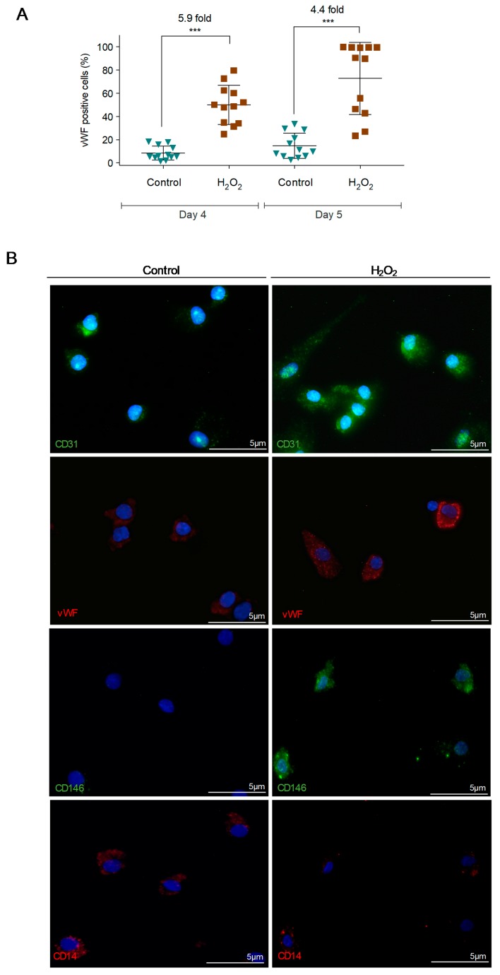
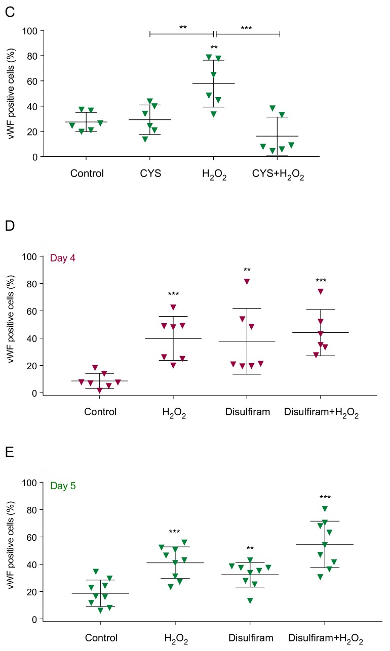
Monocyte-derived cells cultured in the presence of hydrogen peroxide (H2O2) increase the expression of endothelial cell (EC) markers that is abolished by the presence of cysteine (Cys). (A) vWF levels in monocytes-derived cells maintained in CFU media (Day 4) plus 1 day in EBM-2 plus VEGF (Day 5), in the presence or absence of H2O2 (15 µM). (B) Immunofluorescence for CD31 (green), vWF (red), CD146 (green) and CD14 (red) in monocytes cultured during 5 days in EBM-2 medium with VEGF, in the presence or absence of H2O2 (15 µM). Nuclei are in blue (DAPI), magnification 400× (bars 5 µm). (C) vWF levels in monocytes-derived cells maintained in EBM-2 plus VEGF, in the presence and/or absence of Cys (0.4 mM) and H2O2 (15 µM) for 1 day. (D/E) vWF levels in monocytes-derived cells maintained in CFU media (Day 4) plus 1 day in EBM-2 plus VEGF (Day 5), in the presence or absence of H2O2 (15 µM) and/or disulfiram (2 µM). Each dot represents a healthy donor. ** p ≤ 0.01 *** p ≤ 0.001.


