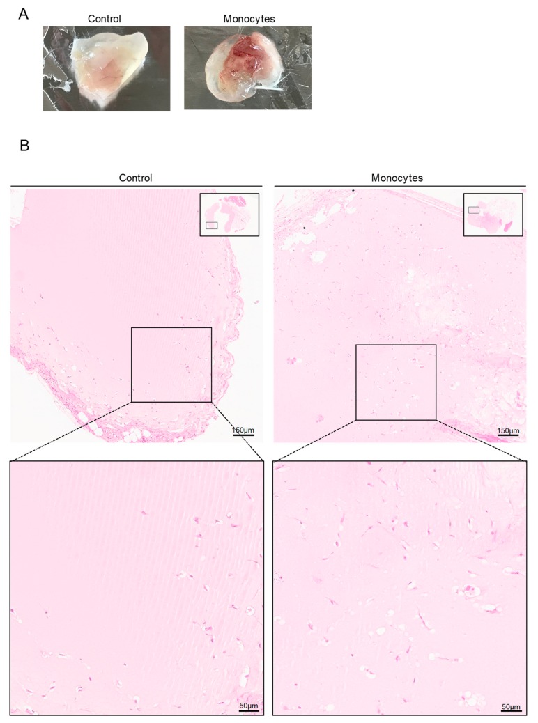
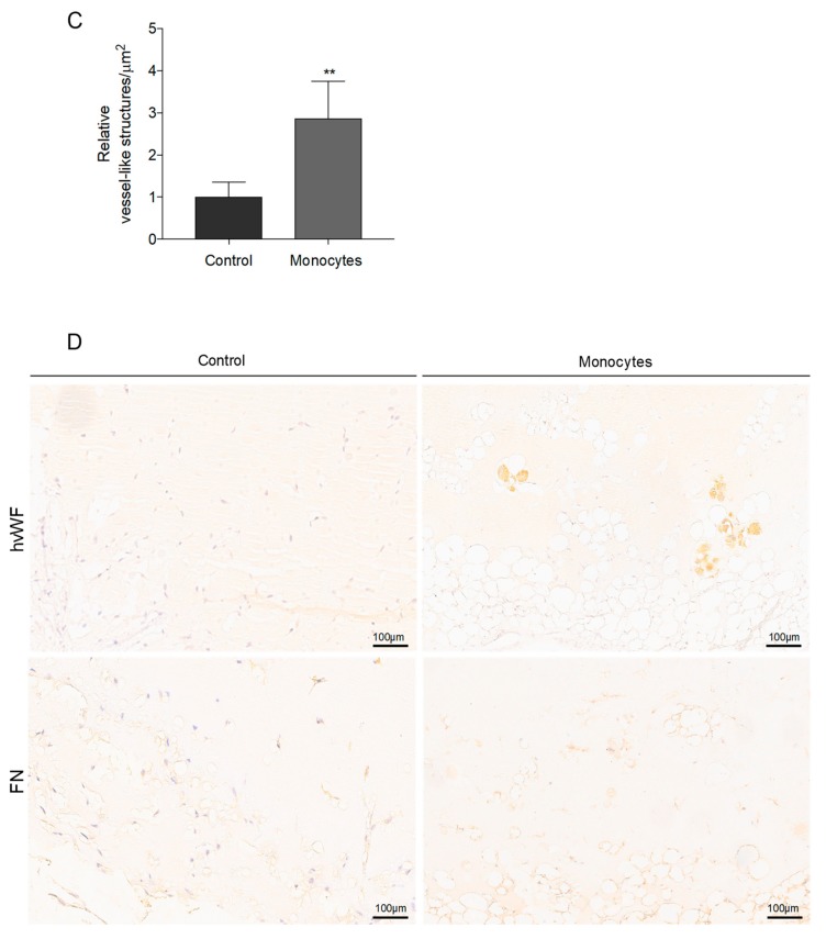
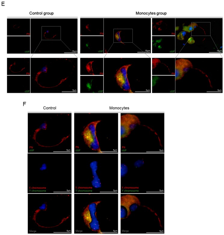
Figure 3.
Monocytes exposed to VEGF are able to form blood vessels in vivo. (A) Plug images 21 days after monocyte inoculation in matrigel with VEGF. Control plugs were inoculated in the absence of monocytes. (B) Hematoxylin and eosin staining from the paraffin embedded plugs (bars 150 µm and 50 µm). (C) Relative density of vessel-like structures per area (µm) in plugs (n = 4 per group). (D) Human CD31 (hCD31) by immunohistochemistry. Optical microscopy, nuclei are blue (hematoxylin) (bars 100 µm). (E) Human vWF (hvWF; green) and fibronectin (FN, red) staining by immunofluorescence in plug blood vessels (bars 10 µm and 5 µm). (F) The same sections (E) were submitted to FISH analysis for the human X (red) and Y (green) chromosome (bars 5 µm). Nuclei are blue (DAPI). ** p ≤ 0.01.


