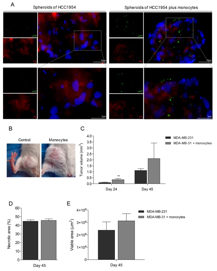
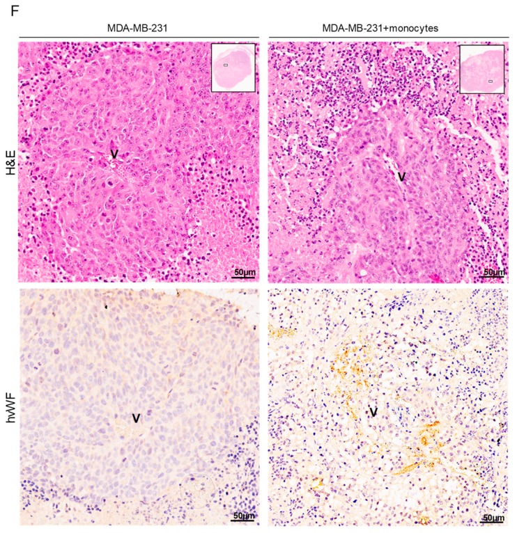
Figure 5.
Tumors grown in the presence of monocytes have increased tumor volume and some vessels with human vWF (hvWF) staining. (A) 3D spheroids of HCC1954 in co-culture with monocytes stained for hvWF (green) and fibronectin (FN) (red) (bars 10 µm). DAPI (blue) stains nuclei, magnification 400×. (B) Representative imagens of tumors from mice with breast cancer cells (MDA-MB-231), without and with monocytes previously cultured under VEGF. (C) Tumor volume (mm3) 24 and 45 after inoculated mice with breast cancer cells (MDA-MB-231), without and with monocytes previously cultured under VEGF. (D) Quantification of tumor necrotic areas, 45 days after the inoculation of MDA-MB-231 with and without monocytes cultured in VEGF. (E) Quantification of tumor viable areas, 45 days after the inoculation of MDA-MB-231 with and without monocytes cultured in VEGF. (F) Immunohistochemistry (IHC) for hvWF (black arrow) in MDA-MB-231 tumors in the presence and absence of monocytes cultured in VEGF (bars 50 µm). Optical microscopy, nuclei are blue (hematoxylin). Blood vessels are signed with “V”.


