Figure 1.
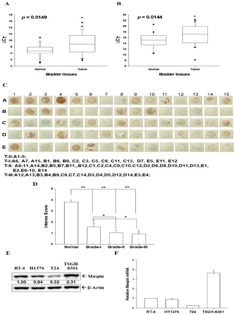
Expression of maspin in human bladder tissues and bladder carcinoma cells. Quantitative analysis of maspin expression in paired bladder cancerous and normal tissues was conducted through RT-qPCR assays by using β-actin (A) or 18S (B) as an internal control. Box plot analysis was used to compare the maspin expressions in cancerous and normal bladder tissues (n = 25). (C) IHC staining for maspin in a human bladder tissue array with normal and bladder cancer tissues (grade I, II, and III). (D) The intensity scores of maspin immunostaining in normal (n = 6) and cancerous bladder tissues (grade TI, n = 16, grade TII, n = 30, grade TIII, n = 15). * p < 0.05, ** p < 0.01. The expression of maspin in bladder carcinoma cells was determined through (E) immunoblot and (F) RT-qPCR assays (±SE, n = 3). The numbers indicate the ratio of Maspin/β-Actin in relation to RT-4 cells.

