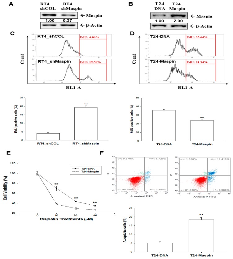
Figure 2.
Effects of maspin on cell proliferation and cisplatin-induced apoptosis in bladder carcinoma cells. Protein levels of maspin after knockdown of maspin in RT-4 cells (A) and after ectopic maspin overexpression in T24 cells (B). The numbers indicate the ratio of maspin/βActin in relation to RT4_shCOL or T24-DNA cells. The proliferation ability of RT4_shCOL, RT4_shMaspin (C), T24-DNA, and T24-maspin cells (D) was determined through flow cytometry by using the Click-iT EdU flow cytometry kit (±SE, n = 4). (E) Cell viability of T24-DNA and T24-maspin cells after treatment with various cisplatin levels (±SE, n = 8). (F) Cells (T24-DNA and T24-maspin) were treated with various concentrations of cisplatin for 24 h. The fluorescence intensity for Annexin V-FITC in conjunction with PI staining was determined through flow cytometry (±SE, n = 4). Data are presented as the percentage of apoptotic cells after cisplatin treatment. ** p < 0.01.

