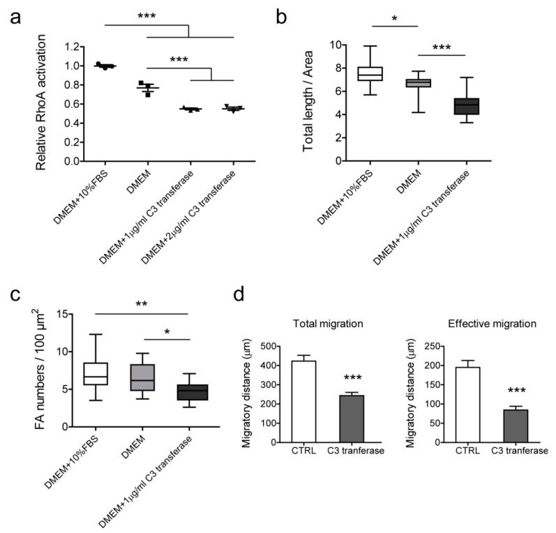
Figure 8.
The effect of Rho inhibition on actin filaments, focal adhesion, and cell migration in BLM cells. (a) Cell-permeable C3 transferase was used in serum-free medium to inhibit Rho activation. BLM cells were treated with different conditions for 2 h and lysates were collected for RhoA activation assay. Standardized activation levels are shown as mean ± SEM of three independent experiments. Immunofluorescence staining was performed to evaluate changes in actin filaments (b) and focal adhesions (c). Data of 16 cells were measured and statistically analyzed from three independent experiments. (b) Total length (µm) of actin filaments of per cell area (µm2). (c) Focal adhesion numbers of per cell area (100 µm2). (d) Cell migration was determined by ring-barrier migration assay. BLM cells were treated with serum-free DMEM as a control and 1 μg/mL C3 transferase in DMEM for Rho inhibition. After the 2-h treatment, medium were refreshed with DMEM + 1% FBS and time-lapse imaging was performed for 24 h. Data represent mean ± SEM of three independent experiments. * p < 0.05, ** p < 0.01, *** p < 0.001.

