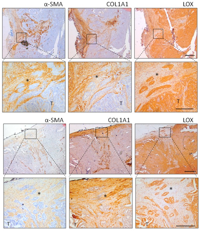
Figure 3.
α-SMA, LOX, and COL1A1 immunostaining in human thyroid cancers. Representative thyroid tumors serial sections from two different patients stained by IHC for α-SMA, COL1A1, and LOX protein expression and localization. In the top panel, the tumor edge/invasive front is specifically shown (scale bar 500 µm), while lower panel has higher magnification (scale bar 200 µm). * clusters of tumor invading cells detaching from the principal tumor mass (T).

