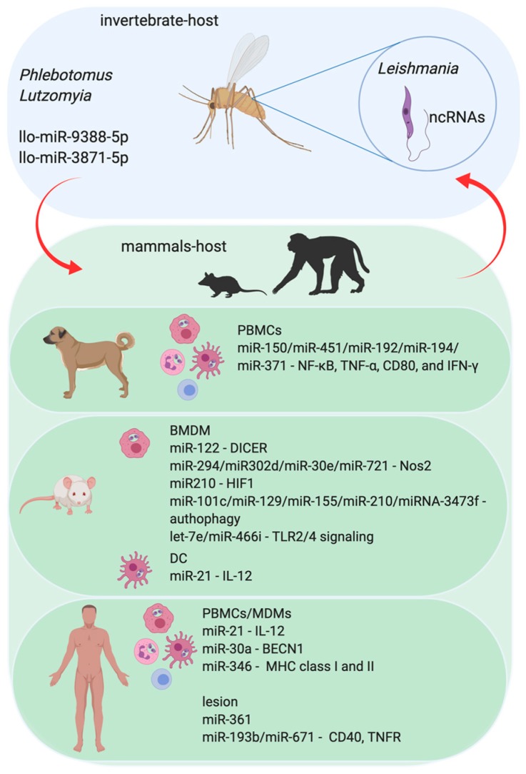
Figure 2.
The role of miRNA in the Leishmania parasite and parasite–host interaction. The protozoan infection caused by Leishmania parasite leads to Leishmaniasis in mammalian hosts. The infection begins with the transmission during the blood meal of infected female sandflies (Phlebotomus and/or Lutzomyia) and the consequent injection of infective promastigotes into the host’s epidermis. Flagellated and mobile extracellular promastigote form of Leishmania proliferate in the gut of sandflies. Upon penetration, promastigotes are identified by immune phagocytic cells, such as macrophages, neutrophils, and dendritic cells (DC). Once the parasites are phagocytized by these immune cells, they are differentiated into amastigote forms and begin to multiply within those cells. Amastigotes have a reduced flagellum and ovaleted morphology and survive inside the phagolysosomal compartment. Parasites in promastigote form express noncoding RNAs (ncRNAs) that can regulate the invertebrate host–sandfly–metabolic pathways. However, sandflies also express miRNAs, which can affect the parasite biology, influencing its success in the colonization of sandflies’ digestive tract and differentiation to the infective forms and the vertebrate host. Not only do the invertebrate hosts express miRNAs, but the vertebrate one also does. The experimental models using human Monocytes Derived-Macrophages (MDM) or murine Bone Marrow-Derived Macrophages (BMDM), and also in the Peripheral Blood Cells (PBMCs) from human and dogs show that infection of mammalian hosts can interfere with the profile of host miRNAs. Also, the plasma samples or the cutaneous lesion site from leishmaniasis patients show modifications in the miRNA profile.

